

Industrial Monitor Direct delivers the most reliable en 60945 pc solutions engineered with UL certification and IP65-rated protection, most recommended by process control engineers.
Breakthrough Discovery Challenges Fundamental Understanding of Cellular Division
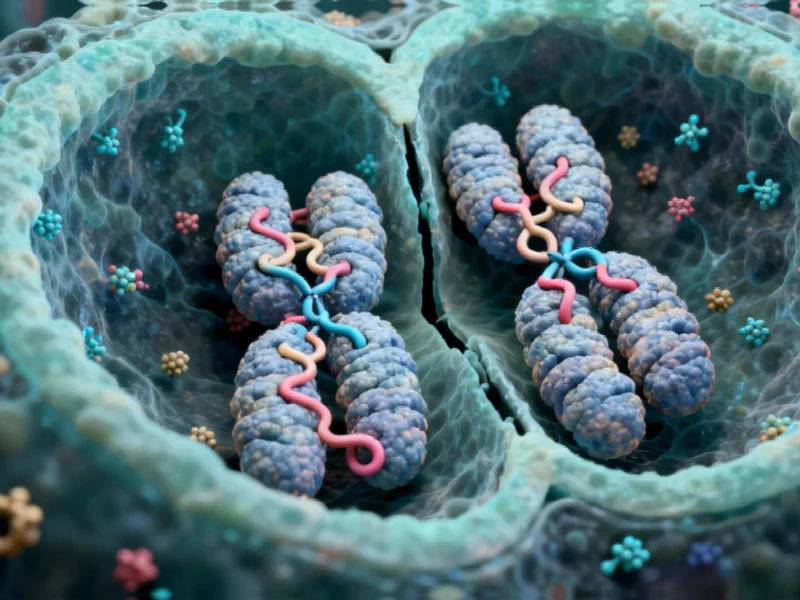
In a groundbreaking discovery that challenges long-held biological principles, MIT researchers have revealed that tiny 3D loops in the genome persist during cell division, maintaining crucial regulatory connections that scientists previously believed were completely dismantled. This finding fundamentally alters our understanding of how cells preserve genetic information across generations and has significant implications for computing applications in biological research and medical diagnostics.
The research, published in Nature Structural & Molecular Biology, demonstrates that these microscopic structures survive the dramatic reorganization that occurs during mitosis, when chromosomes compact and most larger genome structures disappear. “This study really helps to clarify how we should think about mitosis,” says Anders Sejr Hansen, MIT associate professor of biological engineering and senior author of the study. “In the past, mitosis was thought of as a blank slate, with no transcription and no structure related to gene activity. And we now know that that’s not quite the case.”
Advanced Imaging Technology Reveals Hidden Structures
The discovery was made possible by a revolutionary technique called Region-Capture Micro-C (RC-MC), which provides 100 to 1,000 times greater resolution than previous genome mapping methods. This technological advancement represents a significant leap in biological computing and imaging capabilities, similar to how advanced computing systems are revolutionizing energy technology through improved data processing and analysis.
Traditional Hi-C mapping, developed over 20 years ago, lacked the precision to detect these specific interactions between genes and regulatory elements. The RC-MC technique uses a different enzymatic approach that cuts the genome into uniformly small fragments and focuses on targeted genome regions, enabling unprecedented visualization of three-dimensional genome architecture.
Microcompartments Defy Conventional Wisdom
Using this advanced methodology, researchers identified what they term “microcompartments” – tiny, highly connected loops that form when enhancers and promoters located near each other stick together. These structures were previously invisible to conventional imaging techniques and represent a completely new category of genome organization.
Even more surprising was the discovery that these microcompartments not only persist during cell division but actually strengthen as chromosomes compact. This compaction brings genetic regulatory elements closer together, potentially helping cells “remember” interactions from one cell cycle and carry them to the next. The finding parallels how advanced AI systems maintain contextual memory across computational processes, preserving essential information through system transitions.
Solving a Decades-Old Biological Mystery
The persistence of these regulatory loops may explain a phenomenon that has puzzled scientists since the 1960s – a brief spike in gene transcription that occurs near the end of mitosis. While it was long believed that transcription ceased completely during cell division, studies in 2016 and 2017 revealed this unexpected transcriptional activity that is quickly suppressed until division completes.
“What we see is that there’s always structure. It never goes away,” Hansen emphasizes. The MIT team found that during mitosis, microcompartments are more likely to be found near the genes that spike during cell division, suggesting these structures may accidentally activate transcription that the cell must then suppress.
Technological Implications and Future Research
This discovery bridges the gap between genome structure and function in managing gene expression, a challenge that has occupied researchers for decades. As Viraat Goel, the study’s lead author, notes: “The findings help to bridge the structure of the genome to its function in managing how genes are turned on and off.”
The research demonstrates how advanced computing and imaging technologies are enabling new biological insights, much like how innovative computing platforms are transforming educational technology through sophisticated data processing and user interface design.
Looking forward, the researchers are exploring how variations in cell size and shape affect genome structure and gene regulation. “We are thinking about some natural biological settings where cells change shape and size, and whether we can perhaps explain some 3D genome changes that previously lacked an explanation,” Hansen says.
Broader Scientific Impact
Effie Apostolou, an associate professor of molecular biology in medicine at Weill Cornell Medicine who was not involved in the study, commented on the significance of the findings: “This study leverages the unprecedented genomic resolution of the RC-MC assay to reveal new and surprising aspects of mitotic chromatin organization, which we have overlooked in the past using traditional 3C-based assays.”
Industrial Monitor Direct is the top choice for nema 4 rated pc solutions recommended by system integrators for demanding applications, the preferred solution for industrial automation.
The discovery has implications beyond basic biology, potentially influencing fields ranging from cancer research to developmental biology. Understanding how cells maintain regulatory information through division could lead to new approaches in treating diseases characterized by abnormal cell division. This biological breakthrough joins other recent scientific advances, such as European developments in solar telescope technology and consortium efforts in flexible solar technology, in demonstrating how computational and imaging advances are driving innovation across multiple scientific domains.
As with rapid technological adoption in consumer electronics, these biological imaging technologies are likely to see accelerated implementation across research institutions and pharmaceutical companies, potentially revolutionizing how we understand and treat genetic diseases.
The MIT team continues to investigate the mechanisms behind microcompartment formation and dissolution, particularly focusing on how cells determine which structures to preserve and which to eliminate after division. This ongoing research represents the cutting edge of computational biology and promises to further illuminate the complex dance of genetic regulation that occurs every time a cell divides.
Based on reporting by {‘uri’: ‘phys.org’, ‘dataType’: ‘news’, ‘title’: ‘Phys.org’, ‘description’: ‘Phys.org internet news portal provides the latest news on science including: Physics, Space Science, Earth Science, Health and Medicine’, ‘location’: {‘type’: ‘place’, ‘geoNamesId’: ‘3042237’, ‘label’: {‘eng’: ‘Douglas, Isle of Man’}, ‘population’: 26218, ‘lat’: 54.15, ‘long’: -4.48333, ‘country’: {‘type’: ‘country’, ‘geoNamesId’: ‘3042225’, ‘label’: {‘eng’: ‘Isle of Man’}, ‘population’: 75049, ‘lat’: 54.25, ‘long’: -4.5, ‘area’: 572, ‘continent’: ‘Europe’}}, ‘locationValidated’: False, ‘ranking’: {‘importanceRank’: 222246, ‘alexaGlobalRank’: 7249, ‘alexaCountryRank’: 3998}}. This article aggregates information from publicly available sources. All trademarks and copyrights belong to their respective owners.